Pelvic Anatomy Muscles - Pelvis And Perineum Anatomy Vessels Nerves Kenhub - The diaphragm is an essential partner with the pelvic floor, creating the top of the abdominal canister while the pelvic floor forms the bottom.. The pubococcygeus (pc) muscle is the muscle that runs the show in pelvic floor health. The diaphragm is an essential partner with the pelvic floor, creating the top of the abdominal canister while the pelvic floor forms the bottom. A proper kegel exercise means a full contraction and relaxation of the pc muscle. The pelvic floor muscles provide foundational support for the intestines and bladder. The labeled structures are (excluding the correct side):
The levator ani muscles consist of three. The pelvic floor is a group of 16 muscles that work together as a functional unit to provide support, control, sexual function and stability to the pelvis and spine. A proper kegel exercise means a full contraction and relaxation of the pc muscle. Provide individual instruction in exercise performance. Gluteal region muscles that move the femur most muscles that insert on the femur (the thigh bone) and move it, originate on the pelvic girdle.
Pelvic Floor Disorders Anatomy Primal Pictures from www.primalpictures.com Some of the largest and most powerful muscles in the body are the gluteal muscles or gluteal group. The levator ani muscles are the largest group of muscles in the pelvis. The levator ani muscles consist of three. The psoas major and iliacus make up the iliopsoas group. The main function of the pelvic floor muscles are: Individualize the educational component along with exercise performance. Muscles the muscles of the pelvic floor are collectively referred to as the levator ani and coccygeus muscles. The muscles of the pelvis form its floor.
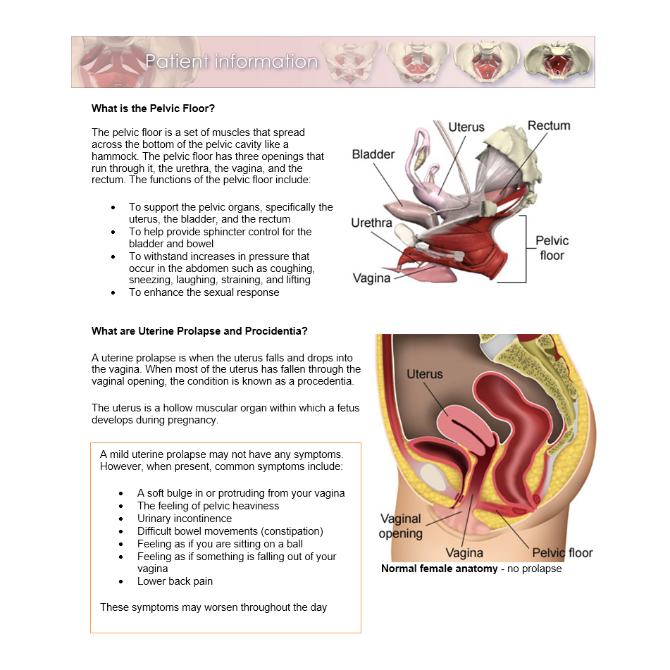
They have several functions, including helping to support the pelvic organs.
Ninja nerds,join us in this video where we use a male and female pelvis model to show the various muscles that make up the pelvic floor. The pelvic diaphragm is a wide but thin muscular layer of tissue that forms the inferior border of the abdominopelvic cavity. It connects the axial skeleton to the lower limbs. The pelvis's frame is made up of the bones of the pelvis, which connect the axial skeleton to the femurs, and therefore acts in weight bearing of the upper body. An important group of muscles in the pelvis is the pelvic floor. Gluteal region muscles that move the femur most muscles that insert on the femur (the thigh bone) and move it, originate on the pelvic girdle. The pelvic floor muscles provide foundational support for the intestines and bladder. Ascending colon superior mesenteric vein superior mesenteric artery gonadal vessels linea semilunaris abdominal aorta linea alba inferior vena cava inferior mesenteric artery infe. The pelvic floor is a group of 16 muscles that work together as a functional unit to provide support, control, sexual function and stability to the pelvis and spine. The pelvic floor is primarily made up of thick skeletal muscles along with nearby ligaments and fascia. The floor of the pelvis is made up of the muscles of the pelvis, which support its contents and maintain urinary and faecal continence. Pelvic muscles that cross the hip joint and attach onto the thigh/leg muscles that cross the hip joint are usually thought of with respect to their open chain motion of the thigh relative to the pelvis at the hip joint. 6.1a, b) is a bony ring consisting of paired innominate bones, the sacrum and coccyx.the innominate bones articulate with each other anteriorly and with the sacrum posteriorly.
The pubococcygeus (pc) muscle is the muscle that runs the show in pelvic floor health. There are many muscles that form the pelvic floor, including puborectalis, pubococcygeus, iliococcygeus and coccygeus. Attached to the pelvis are muscles of the buttocks, the lower back, and the thighs. The main function of the pelvic floor muscles are: It originates from the ischial spines and travels to the lateral aspect of the sacrum and coccyx, along the sacrospinous ligament.
Working Your Pelvic Floor The Pelvic Floor Pelvic Floor First from www.pelvicfloorfirst.org.au These muscles are particularly responsible for the support of pelvic organs and maintenance of continence. Created by physicians for you to help you understand the pelvic floor. Helps improve kegel and pelvic floor. This mri male pelvis axial cross sectional anatomy tool is absolutely free to use. The pelvic floor is a group of 16 muscles that work together as a functional unit to provide support, control, sexual function and stability to the pelvis and spine. The pelvic diaphragm is a wide but thin muscular layer of tissue that forms the inferior border of the abdominopelvic cavity. The iliacus muscle is part of a complex muscle system in the hip area that can function on its own or with other muscles. The deepest layer is the pelvic diaphragm, the muscles that make up the pelvic diaphragm are pubococcygeus, puborectalis, pubourethralis, iliococcygeus and ischiococcygeus.
Teach the patient about pelvic floor anatomy and function.
It is located in the superficial perineal pouch of the pelvic floor, and is not visible externally. Attached to the pelvis are muscles of the buttocks, the lower back, and the thighs. Pelvic floor anatomy that is easy to understand! It originates from the ischial spines and travels to the lateral aspect of the sacrum and coccyx, along the sacrospinous ligament. The floor of the pelvis is made up of the muscles of the pelvis, which support its contents and maintain urinary and faecal continence. These muscles, including the gluteus maximus and the hamstrings, extend the thigh at the hip in support of the body's weight and propulsion. The pelvic diaphragm is a wide but thin muscular layer of tissue that forms the inferior border of the abdominopelvic cavity. The figure above shows the pelvic floor muscles, the iliac muscle, and the muscles that originate from the lumbar vertebrae and attach to the dorsal edge of the ilium As such, you can also divide the musculature that moves the thigh at the hip joint into quadrants. The pelvic floor is a group of 16 muscles that work together as a functional unit to provide support, control, sexual function and stability to the pelvis and spine. A proper kegel exercise means a full contraction and relaxation of the pc muscle. Gluteal region muscles that move the femur most muscles that insert on the femur (the thigh bone) and move it, originate on the pelvic girdle. Ebraheim's educational animated video describes the anatomy of the pelvis, the bony structures, ligaments, muscles, blood supply, and nerves.this video a.
Ascending colon superior mesenteric vein superior mesenteric artery gonadal vessels linea semilunaris abdominal aorta linea alba inferior vena cava inferior mesenteric artery infe. These muscles, including the gluteus maximus and the hamstrings, extend the thigh at the hip in support of the body's weight and propulsion. The pelvis also houses the reproductive organs, which have their own muscles. It connects the axial skeleton to the lower limbs. As such, you can also divide the musculature that moves the thigh at the hip joint into quadrants.
Pelvis Hip Anatomy from uploads-ssl.webflow.com Ebraheim's educational animated video describes the anatomy of the pelvis, the bony structures, ligaments, muscles, blood supply, and nerves.this video a. The pelvic floor is a group of 16 muscles that work together as a functional unit to provide support, control, sexual function and stability to the pelvis and spine. Provide individual instruction in exercise performance. The muscles of the pelvis form its floor. The main function of the pelvic floor muscles are: The pubococcygeus (pc) muscle is the muscle that runs the show in pelvic floor health. These muscles are particularly responsible for the support of pelvic organs and maintenance of continence. Use the mouse scroll wheel to move the images up and down alternatively use the tiny arrows (>>) on both side of the image to move the images.>>) on both side of the image to move the images.
The iliacus muscle is part of a complex muscle system in the hip area that can function on its own or with other muscles.
The iliacus muscle is part of a complex muscle system in the hip area that can function on its own or with other muscles. Teach the patient about pelvic floor anatomy and function. 6.1a, b) is a bony ring consisting of paired innominate bones, the sacrum and coccyx.the innominate bones articulate with each other anteriorly and with the sacrum posteriorly. It is located in the superficial perineal pouch of the pelvic floor, and is not visible externally. The pelvis also houses the reproductive organs, which have their own muscles. They form a large sheet of skeletal muscle that is thicker in some areas than in others. They have several functions, including helping to support the pelvic organs. There are many muscles that form the pelvic floor, including puborectalis, pubococcygeus, iliococcygeus and coccygeus. As such, you can also divide the musculature that moves the thigh at the hip joint into quadrants. All dimensions of muscle complex with visual aids for better understanding of anatomy and functions of pelvic floor muscle. The pelvic diaphragm is a wide but thin muscular layer of tissue that forms the inferior border of the abdominopelvic cavity. Attached to the pelvis are muscles of the buttocks, the lower back, and the thighs. Ascending colon superior mesenteric vein superior mesenteric artery gonadal vessels linea semilunaris abdominal aorta linea alba inferior vena cava inferior mesenteric artery infe.
The iliacus muscle is part of a complex muscle system in the hip area that can function on its own or with other muscles pelvic anatomy. All dimensions of muscle complex with visual aids for better understanding of anatomy and functions of pelvic floor muscle.
Pelvic Anatomy Muscles - Pelvis And Perineum Anatomy Vessels Nerves Kenhub - The diaphragm is an essential partner with the pelvic floor, creating the top of the abdominal canister while the pelvic floor forms the bottom.. There are any Pelvic Anatomy Muscles - Pelvis And Perineum Anatomy Vessels Nerves Kenhub - The diaphragm is an essential partner with the pelvic floor, creating the top of the abdominal canister while the pelvic floor forms the bottom. in here.